■サンプル写真(詳細)■
対物5x,10x,20x,40x、撮影レンズ3.3xで撮影しています
標本が大きく対物5xでも全体が入らないもの
標本が小さく対物40xでは撮影出来ないもの
があります
予めご了承ください

001―Pig Adipose Cell (whole mount)【顕微鏡プレパラート標本】
【豚 - 脂肪細胞(全組織標本)】

002―Mouse Cuboidal Epithelium (section)【顕微鏡プレパラート標本】
【マウス - 立方上皮(切片)】

003―Dog Columnar Epithelium(section)【顕微鏡プレパラート標本】
【犬 - 円柱上皮(切片)】

004―Dog Columnar Ciliated Epithelium (section)【顕微鏡プレパラート標本】
【犬 - 円柱線毛上皮(切片)】

005―Paddy Pollen (whole mount)【顕微鏡プレパラート標本】
【稲花粉(全組織標本)】

006―Fresh Water Fish Gill (section)【顕微鏡プレパラート標本】
【淡水魚えら(切片)】

007―Hydra (longitudinal section)【顕微鏡プレパラート標本】
【ヒドラ(縦断)】

008―Hydra Plain and Budding (whole mount)【顕微鏡プレパラート標本】
【ヒドラ - 出芽(全組織標本)】

009―Planaria (cross section)【顕微鏡プレパラート標本】
【プラナリア(横断)】

010―Blood Fluke - Eggs (whole mount)【顕微鏡プレパラート標本】
【住血吸虫 - 卵(全組織標本)】

011―Taenia Pisiformis (whole mount)【顕微鏡プレパラート標本】
【豆状条虫(全組織標本)】

012―Ascaris (cross section)【顕微鏡プレパラート標本】
【回虫(横断)】

013―Mosquito-Female (whole mount)【顕微鏡プレパラート標本】
【蚊 - メス(全組織標本)】

014―Mitosis-Acaris Egg(cross section)【顕微鏡プレパラート標本】
【ダニの卵 - 細胞分裂(横断)】

015―Mantis Leg (whole mount)【顕微鏡プレパラート標本】
【カマキリ - 足(全組織標本)】

016―Dog Tongue (longitudinal section)【顕微鏡プレパラート標本】
【犬 - 舌(縦断)】

017―Dog Stomach Cardiac Region (section)【顕微鏡プレパラート標本】
【犬 - 胃噴門(切片)】

018―Dog Stomach Pyloric Region (section)【顕微鏡プレパラート標本】
【犬 - 胃幽門(切片)】

019―Pig Liver (section)【顕微鏡プレパラート標本】
【ブタ - 肝臓(切片)】

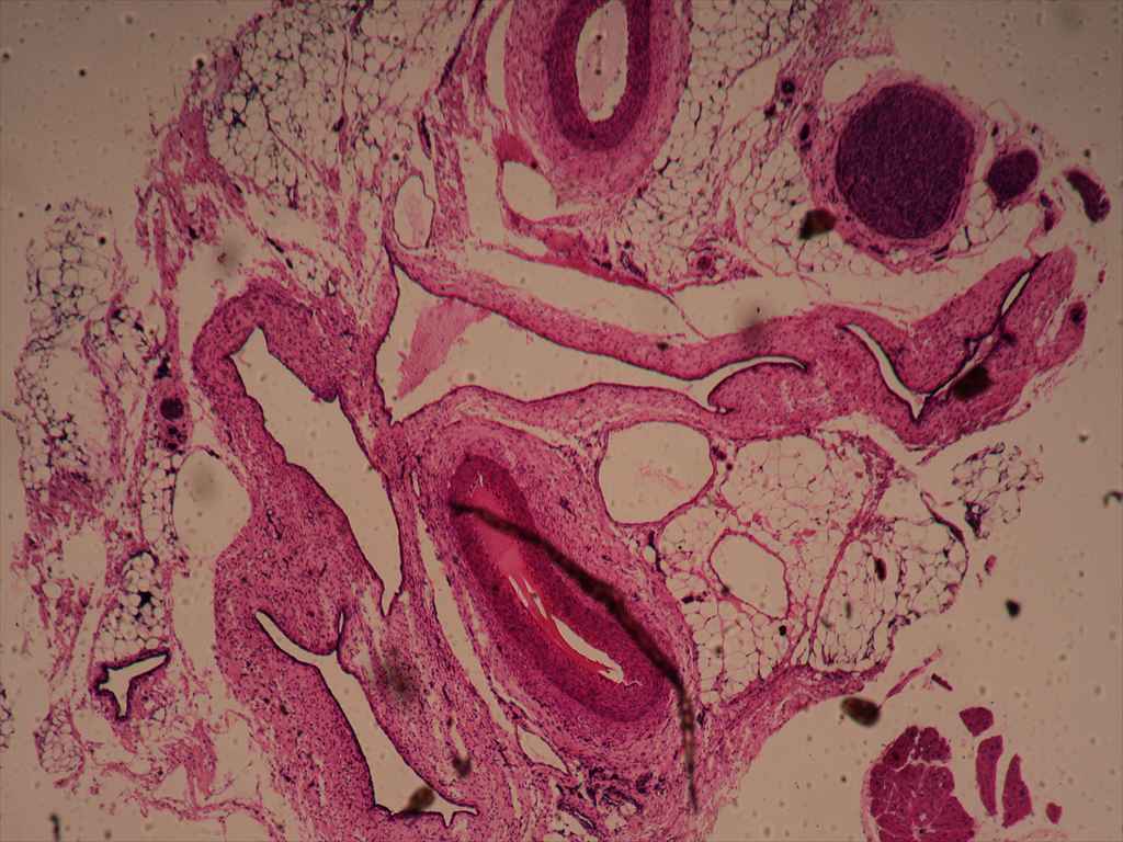
020―Rabbit Venae Cavac (cross section)【顕微鏡プレパラート標本】
【ウサギ - 大静脈(横断)】

021―Mouse Kidney (section)【顕微鏡プレパラート標本】
【マウス - 腎臓(切片)】

022―Dog Urinary Bladder (section)【顕微鏡プレパラート標本】
【犬 - 膀胱(切片)】

023―Human Skin - Hair - Follicle (section)【顕微鏡プレパラート標本】
【人 - 皮膚 - 髪 - 毛包(切片)】

024―Rabbit Ganglia (section)【顕微鏡プレパラート標本】
【ウサギ - 神経節(切片)】

025―Dog Olfactory Membrane (section)【顕微鏡プレパラート標本】
【犬 - 嗅膜(切片)】

026―Mouse Ovary (section)【顕微鏡プレパラート標本】
【マウス - 卵巣(切片)】

027―Frog Spermatozoa (smear)【顕微鏡プレパラート標本】
【カエル - 精子(塗抹標本)】

028―Bulgarious (smear)【顕微鏡プレパラート標本】
【乳酸菌(塗抹標本)】

029―Lichen (section)【顕微鏡プレパラート標本】
【地衣類(切片)】

030―Maroharttis Archegortia (longitudinal section)【顕微鏡プレパラート標本】
【ゼニゴケ - 造卵器(縦断)】

031―Marchantia Aritheridis (longitudinal section)【顕微鏡プレパラート標本】
【ゼニゴケ - 造精器(縦断)】

032―Polytrichum (whole mount)【顕微鏡プレパラート標本】
【スギゴケ(全組織標本)】

033―Polytrichum Antheridia (longitudinal section)【顕微鏡プレパラート標本】
【スギゴケ - 造精器(縦断)】

034―Polytrichum Archegonis (longitudinal section)【顕微鏡プレパラート標本】
【スギゴケ - 造卵器(縦断)】

035―Fern Root (cross section)【顕微鏡プレパラート標本】
【シダ - 根(横断)】

036―Fern Stem (cross section)【顕微鏡プレパラート標本】
【シダ - 幹(横断)】

037―Fern Leaf - Sorus (cross section)【顕微鏡プレパラート標本】
【シダ - 葉 - 胞子嚢群(横断)】

038―Pine Stem (longitudinal section)【顕微鏡プレパラート標本】
【松 - 幹(縦断)】

039―Pine Cone-Female(longitudinal section)【顕微鏡プレパラート標本】
【松 - 雌花(縦断)】

040―Vicia Faba Root, Tip (longitudinal section)【顕微鏡プレパラート標本】
【ソラマメ - 根端(縦断)】

041―Ranunculus Root (cross section)【顕微鏡プレパラート標本】
【キンポウゲ - 根(横断)】

042―Pisum Seed (longitudinal section)【顕微鏡プレパラート標本】
【エンドウ - 種子(縦断)】

043―Stem - Cork Cell (cross section)【顕微鏡プレパラート標本】
【茎 - コルク細胞(横断)】

044―Nymphaea(Spongy tissue)(cross section)【顕微鏡プレパラート標本】
【すいれん(海綿状組織)(横断)】

045―Nymphaea,of Aqustio Stem(cross section)【顕微鏡プレパラート標本】
【すいれんの茎(横断)】

046―Pelargonium, of Leaf (cross section)【顕微鏡プレパラート標本】
【ゼラニウム - 葉(横断)】

047―Striated Muscle (cross section)【顕微鏡プレパラート標本】
【横紋筋(横断)】

048―Stem-Sieve Tube and Companion Cell (longitudinal section)【顕微鏡プレパラート標本】
【茎 - 篩管と伴細胞(縦断)】

049―Root Hair (cross section)【顕微鏡プレパラート標本】
【根毛(横断)】

050―Dense Connective Tissue (section)【顕微鏡プレパラート標本】
【密性結合組織(切片)】

051―Loose Connective Tissue (section)【顕微鏡プレパラート標本】
【疎性結合組織(切片)】

052―Aspergillus (whole mount)【顕微鏡プレパラート標本】
【コウジカビ(全組織標本)】

053―Lung-Injected (Rabbit) (section)【顕微鏡プレパラート標本】
【ウサギ - 肺注射(切片)】

054―Kidney-Injected (Rabbit) (section)【顕微鏡プレパラート標本】
【ウサギ - 腎注射(切片)】

055―Trematodes- Miracidia (whole mount)【顕微鏡プレパラート標本】
【吸虫 - 幼虫(全組織標本)】

056―Mosquito - Male (whole mount)【顕微鏡プレパラート標本】
【蚊 - オス(全組織標本)】

057―Mosquito Eggs (whole mount)【顕微鏡プレパラート標本】
【蚊 - 卵(全組織標本)】

058―Drosophila - Female (whole mount)【顕微鏡プレパラート標本】
【ショウジョウバエ - メス(全組織標本)】

059―Drosophila - Male (whole mount)【顕微鏡プレパラート標本】
【ショウジョウバエ - オス(全組織標本)】

060―Embryo of Shepherd's (section)【顕微鏡プレパラート標本】
【シェパードの胚(切片)】

061―Drosophila - Chrysalis (whole mount)【顕微鏡プレパラート標本】
【ショウジョウバエ - サナギ(全組織標本)】

062―Frog - Vnsegmented egg (section)【顕微鏡プレパラート標本】
【カエル - 未卵割(切片)】

063―Frog - Holoblastic Cleavage (section)【顕微鏡プレパラート標本】
【カエル - 完全卵割(切片)】

064―Frog - Cleavage (section)【顕微鏡プレパラート標本】
【カエル - 卵割(切片)】

065―Frog - Blastule (section)【顕微鏡プレパラート標本】
【カエル - 胞胚(切片)】

066―Cucurbita (whole mount)【顕微鏡プレパラート標本】
【カボチャ(全組織標本)】

067―Pine Pollen (whole mount)【顕微鏡プレパラート標本】
【松花粉(全組織標本)】

068―Plasmodesmata (section)【顕微鏡プレパラート標本】
【原形質連絡(切片)】

069―Silver Berry Scaly Hair (whole mount)【顕微鏡プレパラート標本】
【グミ鱗毛(全組織標本)】

070―Peacock Feather (whole mount)【顕微鏡プレパラート標本】
【クジャクの羽(全組織標本)】

071―Dandelion Fuzz (whole mount)【顕微鏡プレパラート標本】
【タンポポ縮れ毛(全組織標本)】

072―Feather (whole mount)【顕微鏡プレパラート標本】
【羽毛(全組織標本)】

073―Fibre (whole mount)【顕微鏡プレパラート標本】
【繊維(全組織標本)】

074―House Fly Leg (whole mount)【顕微鏡プレパラート標本】
【イエバエ - 足(全組織標本)】

075―House Fly Wing (whole mount)【顕微鏡プレパラート標本】
【イエバエ - ハネ(全組織標本)】

076―Corn Starch (whole mount)【顕微鏡プレパラート標本】
【コーン - 澱粉(全組織標本)】

077―Butterfly Leg (whole mount)【顕微鏡プレパラート標本】
【蝶 - あし(全組織標本)】

078―Ascarid Egg (whole mount)【顕微鏡プレパラート標本】
【回虫卵(全組織標本)】

079―Leaf of Winter Jasmine (cross section)【顕微鏡プレパラート標本】
【冬ジャスミンの葉(横断)】

080―Cerebrum Mammal (section)【顕微鏡プレパラート標本】
【哺乳類 - 大脳】

081―Cerebellum Mammal (section)【顕微鏡プレパラート標本】
【哺乳類 - 小脳】

082―Ant (whole mount)【顕微鏡プレパラート標本】
【アリ(全組織標本)】

083―Vegetable Pollen (whole mount)【顕微鏡プレパラート標本】
【野菜 - 花粉(全組織標本)】

084―Silkworm Moth Antennae (whole mount)【顕微鏡プレパラート標本】
【カイコ蛾 - 触角(全組織標本)】

085―Fruit (cross section)【顕微鏡プレパラート標本】
【果実(横断)】

086―Peach Worm (whole mount)【顕微鏡プレパラート標本】
【シンクイムシ(全組織標本)】

087―Butterfly Antennae (whole mount)【顕微鏡プレパラート標本】
【蝶 - 触角(全組織標本)】

088―Cotton Worm (whole mount)【顕微鏡プレパラート標本】
【綿虫(全組織標本)】

089―Shrimp - Egg (whole mount)【顕微鏡プレパラート標本】
【エビ - 卵(全組織標本)】

090―Letter "E" (whole mount)【顕微鏡プレパラート標本】
【文字 "E"(全組織標本)】

091―Root Bacteria (cross section)【顕微鏡プレパラート標本】
【菌根菌(横断)】

092―Typical Animal Cell 【顕微鏡プレパラート標本】
【典型的動物細胞】

093―Typical Plant Cell 【顕微鏡プレパラート標本】
【典型的植物細胞】

094―Cotton Leaf (cross section)【顕微鏡プレパラート標本】
【綿の葉(横断)】

095―Cotton Stem (cross section)【顕微鏡プレパラート標本】
【綿の茎(横断)】

096―Young Root of Broad Bean (cross section)【顕微鏡プレパラート標本】
【ソラマメの若い根(横断)】

097―Soya Stem (cross section)【顕微鏡プレパラート標本】
【大豆茎(横断)】

098―Silkworm Moth Larva (whole mount)【顕微鏡プレパラート標本】
【カイコ蛾幼虫(全組織標本)】

099―Pea Pollen (whole mount)【顕微鏡プレパラート標本】
【エンドウ花粉(全組織標本)】

100―Ciliated Epithelium (section)【顕微鏡プレパラート標本】
【線毛上皮(切片)】

101―Human Cell Mucus Membrane (smear)【顕微鏡プレパラート標本】
【人 - 細胞粘液膜(塗抹標本)】

102―Frog Epidermic Cell (section)【顕微鏡プレパラート標本】
【カエル - 表皮細胞(切片)】

103―Dog Squamous Epithelium (whole mount)【顕微鏡プレパラート標本】
【犬 - 扁平上皮(全組織標本)】

104―Paramecium (whole mount)【顕微鏡プレパラート標本】
【ゾウリムシ(全組織標本)】

105―Hydra (cross section)【顕微鏡プレパラート標本】
【ヒドラ(横断)】

106―Earthworm (cross section)【顕微鏡プレパラート標本】
【ミミズ(横断)】

107―Daphina (whole mount)【顕微鏡プレパラート標本】
【ミジンコ(全組織標本)】

108―Mosquito Larva (whole mount)【顕微鏡プレパラート標本】
【蚊 - ボウフラ(全組織標本)】

109―Mosquito Mouth Parts (whole mount)【顕微鏡プレパラート標本】
【蚊 - 口器(全組織標本)】

110―Honeybee Mouth Parts (whole mount)【顕微鏡プレパラート標本】
【ミツバチ - 口器(全組織標本)】

111―Housefly Mouth Parts (whole mount)【顕微鏡プレパラート標本】
【イエバエ - 口器(全組織標本)】

112―Honeybee Worker Leg-Composite (whole mount)【顕微鏡プレパラート標本】
【ミツバチ - 働き蜂の足(全組織標本)】

113―Mosquito Wings (whole mount)【顕微鏡プレパラート標本】
【蚊 - 羽(全組織標本)】

114―Butterfly Wings Ocales (whole mount)【顕微鏡プレパラート標本】
【蝶 - 羽(全組織標本)】

115―Dragonfly Wings (whole mount)【顕微鏡プレパラート標本】
【トンボ - 羽(全組織標本)】

116―Honeybee Wings (whole mount)【顕微鏡プレパラート標本】
【ミツバチ - 羽(全組織標本)】

117―Housefly Compound Eye (whole mount)【顕微鏡プレパラート標本】
【イエバエ - 複眼(全組織標本)】

118―Honeybee Compound Eye (whole mount)【顕微鏡プレパラート標本】
【ミツバチ - 複眼(全組織標本)】

119―Dragonfly Compound Eye (whole mount)【顕微鏡プレパラート標本】
【トンボ - 複眼(全組織標本)】

120―Dog Esophagus (cross section)【顕微鏡プレパラート標本】
【犬 - 食道(横断)】

121―Dog Small Intestine (section)【顕微鏡プレパラート標本】
【犬 - 小腸(切片)】

122―Dog Stomach (section)【顕微鏡プレパラート標本】
【犬 - 胃(切片)】

123―Dog Duodenum (cross section)【顕微鏡プレパラート標本】
【犬 - 十二指腸(横断)】

124―Dog Jejunum (cross section)【顕微鏡プレパラート標本】
【犬 - 空腸(横断)】

125―Dog Ileum (cross section)【顕微鏡プレパラート標本】
【犬 - 回腸(横断)】

126―Dog Rectum (cross section)【顕微鏡プレパラート標本】
【犬 - 直腸(横断)】

127―Dog Spleen (section)【顕微鏡プレパラート標本】
【犬 - 脾臓(切片)】

128―Dog Pancreas (section)【顕微鏡プレパラート標本】
【犬 - 膵臓(切片)】

129―Pig Gall Bladder (section)【顕微鏡プレパラート標本】
【豚 - 胆嚢(切片)】
130―Rabbit Artery and Vein (cross section)【顕微鏡プレパラート標本】
【ウサギ - 動脈と静脈(横断)】

131―Rabbit Arteriole (cross section)【顕微鏡プレパラート標本】
【ウサギ - 細動脈(横断)】

132―Human Blood (smear)【顕微鏡プレパラート標本】
【人 - 血液(塗抹標本)】

133―Frog Blood (smear)【顕微鏡プレパラート標本】
【カエル - 血液(塗抹標本)】

134―Fish Blood (smear)【顕微鏡プレパラート標本】
【魚 - 血液(塗抹標本)】

135―Rabbit Lymph Node (section)【顕微鏡プレパラート標本】
【ウサギ - リンパ節(切片)】

136―Dog Trachea (cross section)【顕微鏡プレパラート標本】
【犬 - 気管(横断)】

137―Dog Ureter (cross section)【顕微鏡プレパラート標本】
【犬 - 尿管(横断)】

138―Human Skin Sweat Gland (section)【顕微鏡プレパラート標本】
【人 - 皮膚汗腺(切片)】

139―Human Hair (whole mount)【顕微鏡プレパラート標本】
【人毛(全組織標本)】

140―Feather (whole mount)【顕微鏡プレパラート標本】
【羽毛(全組織標本)】

141―Fish Scales (whole mount)【顕微鏡プレパラート標本】
【魚鱗(全組織標本)】

142―Rabbit Hyaline Cartilage (section)【顕微鏡プレパラート標本】
【ウサギ - 硝子軟骨(切片)】

143―Dog Skeletal Muscle (longitudinal section) &(cross section)【顕微鏡プレパラート標本】
【犬 - 骨格筋(縦断)(横断)】

144―Dog Smooth Muscle (longitudinal section) &(cross section)【顕微鏡プレパラート標本】
【犬 - 平滑筋(縦断)(横断)】

145―Dog Cardiac Muscle (longitudinal section)【顕微鏡プレパラート標本】
【犬 - 心筋(縦断)】

146―Pig Motor Nerve (whole mount)【顕微鏡プレパラート標本】
【豚 - 運動神経(全組織標本)】

147―Rabbit Spinal Cord (cross section)【顕微鏡プレパラート標本】
【ウサギ - 脊髄(横断)】

148―Dog Taste Buds (section)【顕微鏡プレパラート標本】
【犬 - 味蕾(切片)】

149―Rabbit Testis (section)【顕微鏡プレパラート標本】
【ウサギ - 精巣(切片)】

150―Human Spermatozoa (smear)【顕微鏡プレパラート標本】
【人 - 精子(塗抹標本)】

151―Onion Epidermis (whole mount)【顕微鏡プレパラート標本】
【タマネギ表皮(全組織標本)】

152―Hydrilla Verticillata Leaf (whole mount)【顕微鏡プレパラート標本】
【クロモ (水草) - 葉(全組織標本)】

153―Rhoeo Disolor Leaf (whole mount)【顕微鏡プレパラート標本】
【ムラサキオモト - 葉(全組織標本)】

154―Ipomoea Root (whole mount)【顕微鏡プレパラート標本】
【サツマイモ - 根(全組織標本)】

155―Tomato Flesh (whole mount)【顕微鏡プレパラート標本】
【トマト果肉(全組織標本)】

156―Pome Sclereid (whole mount)【顕微鏡プレパラート標本】
【梨状果厚膜細胞(全組織標本)】

157―Mitosis - Onion Root Tip (longitudinal section)【顕微鏡プレパラート標本】
【細胞分裂 - タマネギの根先端(縦断)】

158―Meiosis-Lillitrm Pollen (whole mount)【顕微鏡プレパラート標本】
【ユリ - 子房減数分裂(全組織標本)】

159―Root-Meristem (longitudinal section)【顕微鏡プレパラート標本】
【根 - 分裂組織(縦断)】

160―Stem-Collenchyma (cross section)【顕微鏡プレパラート標本】
【茎 - 厚角組織(横断)】

161―Stem-Parenchyma (cross section)【顕微鏡プレパラート標本】
【茎 - 柔組織(横断)】

162―Stem-Sclerenchvma (cross section)【顕微鏡プレパラート標本】
【茎 - 厚壁組織(横断)】

163―Stem-Tracheid (longitudinal section)【顕微鏡プレパラート標本】
【茎 - 仮道管(縦断)】

164―Mixed Bacteria (smear)【顕微鏡プレパラート標本】
【混合細菌(塗抹標本)】

165―Coccus (smear)【顕微鏡プレパラート標本】
【球菌(塗抹標本)】

166―Bacillus (smear)【顕微鏡プレパラート標本】
【桿菌(塗抹標本)】

167―Spirllum (smear)【顕微鏡プレパラート標本】
【らせん菌(塗抹標本)】

168―Rhizopus (whole mount)【顕微鏡プレパラート標本】
【クモノスカビ(全組織標本)】

169―Penicillium (whole mount)【顕微鏡プレパラート標本】
【ペニシリウム(全組織標本)】

170―Yeast-Budding (whole mount)【顕微鏡プレパラート標本】
【酵母 - 出芽(全組織標本)】

171―Coprinus Mushroom Set (cross section)【顕微鏡プレパラート標本】
【ひとよたけ(横断)】

172―Chlamydomonas (whole mount)【顕微鏡プレパラート標本】
【クラミドモナス属(全組織標本)】

173―Volvox (whole mount)【顕微鏡プレパラート標本】
【ボルボックス(全組織標本)】

174―Spirogyia Conjugation (whole mount)【顕微鏡プレパラート標本】
【アオミドロ - 結合(全組織標本)】

175―Euglena (whole mount)【顕微鏡プレパラート標本】
【ミドリムシ(全組織標本)】

176―Marchrrtia Mature Sporophyte (longitudinal section)【顕微鏡プレパラート標本】
【ゼニゴケ - 成熟胞子体(縦断)】

177―Fern, Leaf-Sorus (whole mount)【顕微鏡プレパラート標本】
【シダ - 葉・胞子嚢群(全組織標本)】

178―Fern Porthallia (whole mount)【顕微鏡プレパラート標本】
【シダ - 前葉体(全組織標本)】

179―Fern Prothallia and Sporangia (whole mount)【顕微鏡プレパラート標本】
【シダ - 前葉体・胞子嚢(全組織標本)】

180―Pine Root (cross section)【顕微鏡プレパラート標本】
【松 - 根(横断)】

181―Pine Stem (cross section)【顕微鏡プレパラート標本】
【松 - 幹(横断)】

182―Pine Leaf (cross section)【顕微鏡プレパラート標本】
【松 - 葉(横断)】

183―Pine Young Staminate, Cone (longitudinal section)【顕微鏡プレパラート標本】
【松 - 若いまつかさ(縦断)】

184―Zea Root, Tip (longitudinal section)【顕微鏡プレパラート標本】
【トウモロコシ - 根先端(縦断)】

185―Zea Root (cross section)【顕微鏡プレパラート標本】
【トウモロコシ - 根(横断)】

186―Zea Stem (longitudinal section)【顕微鏡プレパラート標本】
【トウモロコシ - 茎(縦断)】

187―Cucurbita Stem (longitudinal section)【顕微鏡プレパラート標本】
【カボチャ属 - 茎(縦断)】

188―Zea Stem (cross section)【顕微鏡プレパラート標本】
【トウモロコシ - 茎(横断)】

189―Rice Stem (cross section)【顕微鏡プレパラート標本】
【稲 - 茎(横断)】

190―Sunflower Stem (cross section)【顕微鏡プレパラート標本】
【ヒマワリ - 茎(横断)】

191―Pumpkin Stem (cross section)【顕微鏡プレパラート標本】
【カボチャ - 茎(横断)】

192―Tilia Stem (cross section)【顕微鏡プレパラート標本】
【シナノキ属 - 茎(横断)】

193―teminal Bud Stem Tip (longitudinal section)【顕微鏡プレパラート標本】
【茎最上部の芽(縦断)】

194―Poa Leaf (cross section)【顕微鏡プレパラート標本】
【イチゴツナギ属 - 葉(横断)】

195―Ipomoea Leaf (cross section)【顕微鏡プレパラート標本】
【サツマイモ - 葉(横断)】

196―Stomatal-Vicia Faba Leaf (whole mount)【顕微鏡プレパラート標本】
【気孔-ソラマメの葉 (全組織標本)】

197―Lillium Ovary (cross section)【顕微鏡プレパラート標本】
【ユリ - 子房(横断)】

198―Lillium Anther (cross section)【顕微鏡プレパラート標本】
【ユリ - 葯(横断)】

199―Pollen Gem (whole mount)【顕微鏡プレパラート標本】
【花粉宝石(全組織標本)】

200―Zea Seed (longitudinal section)【顕微鏡プレパラート標本】
【トウモロコシの種(縦断)】
以上
 |